MRCEM
Core Emergency Ultrasound Level 1 (POCUS) Course in Manchester
This one-day POCUS course in Manchester gives doctors and acute-care clinicians supervised, hands-on practice in the core emergency ultrasound scans: the abdominal aorta, free fluid, focused echocardiography and lung ultrasound. Run by Kung Medics in Levenshulme, it teaches you to optimise images, recognise key abnormalities and decide when a finding should change management. No previous scanning experience is needed, and you leave with a CPD-accredited certificate.
- Runs 9:00am to 5:00pm
- Small-group, hands-on supervised scanning
- Aorta, free fluid, focused echo and lung ultrasound
- Modern ultrasound machines and simulation kit
- Suitable for beginners and those refreshing skills
- Certificate issued; CPD-accredited by The CPD Group
- Relevant to MRCEM and FRCEM preparation
- Employer invoicing available at booking
Course summary
- Qualification
- The CPD Group
- Duration
- 1 day
- Format
- Face-to-face
- Location
- Manchester
- CPD
- 6
- Price
- £375
- Next date
- Monday 26 October 2026
Point-of-care ultrasound training in Manchester for acute clinicians
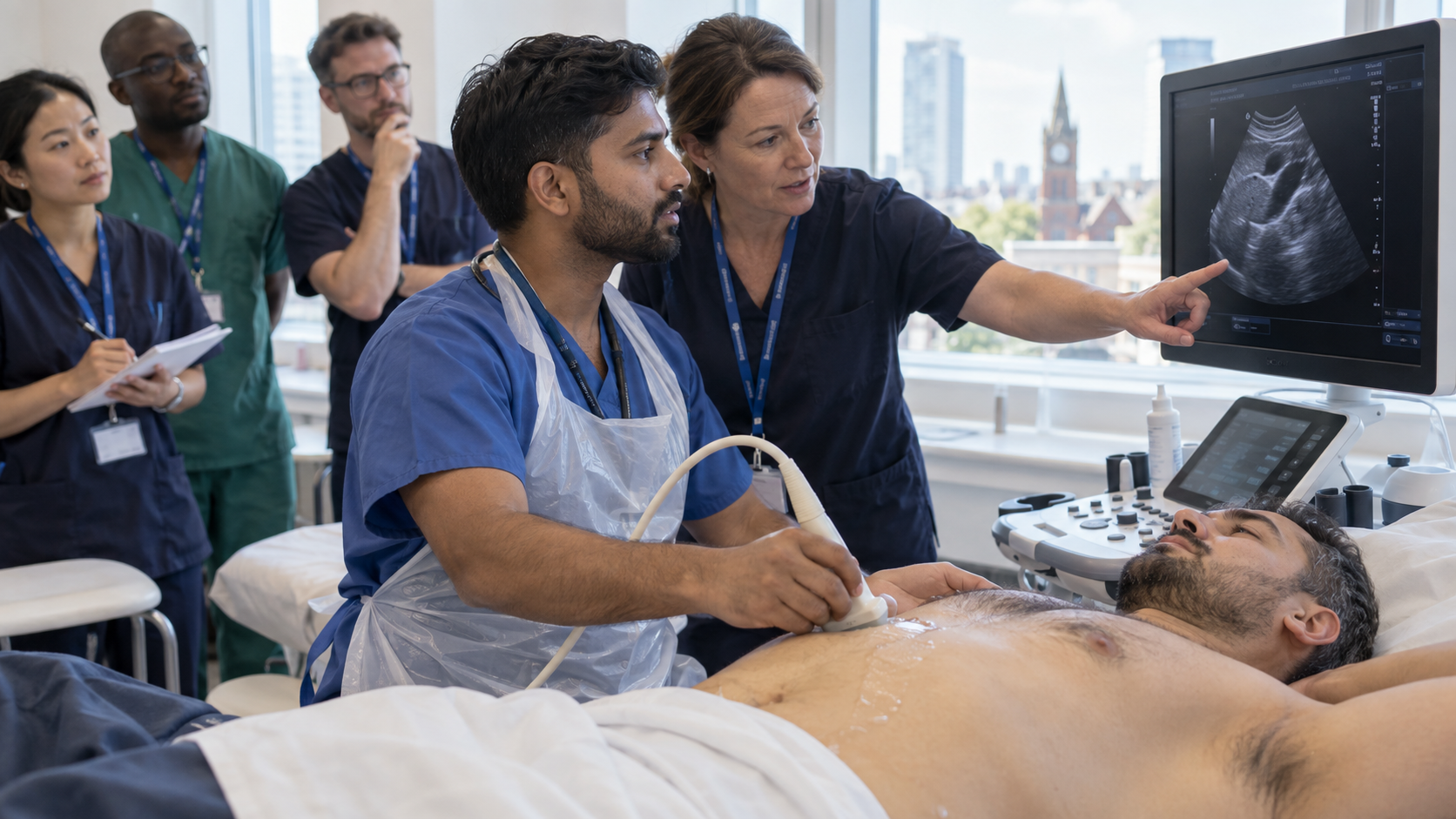
Bedside ultrasound can answer urgent questions in minutes, but only if you can get a usable image and know what it means for the patient in front of you. Our Core Emergency Ultrasound Level 1 POCUS course in Manchester is built around exactly that: a full day of small-group scanning with an instructor beside you, covering the four core applications used in emergency and acute medicine.
Whether you have never held a probe or have sat through ultrasound lectures without the confidence to scan alone, you will spend most of the day with your hands on the machine. Choose a date below to secure your place.
Why learn POCUS with Kung Medics
We teach ultrasound as a clinical tool, not a picture-recognition exercise. Every view is linked to a question you would ask at the bedside of a deteriorating patient: what am I looking for, is this image adequate, and does it change what I do next?
- Scanning first: short explanations, then repeated supervised practice on modern ultrasound machines.
- Small practical groups: instructors spot your individual difficulties and correct technique in real time.
- Simulation: used for repeated needle-guidance practice before you try it clinically.
- Safe practice: clear teaching on limitations, logbooks and local governance so you know how to keep building competence after the day.
Upcoming POCUS course dates in Manchester
£375
Book this dateMore dates
Choosing a date takes you straight to booking with that date selected.
Course overview
The course runs as one full day in Levenshulme, from 9:00am to 5:00pm, and moves from machine basics to integrated clinical scanning.
The day starts with the essentials that make every later scan easier: how ultrasound works, choosing the right probe, orientation and probe markers, and adjusting depth and gain to optimise the image. You then work through the core emergency applications in turn, scanning each one in small groups:
- the abdominal aorta, including telling it apart from the IVC and measuring its diameter
- focused assessment for free fluid in the upper quadrants and pelvis
- focused echocardiography using parasternal, apical and subcostal views
- lung ultrasound for sliding, A-lines, B-lines, effusion and consolidation
Where it is included in the programme, you also practise ultrasound-guided peripheral vascular access on simulation equipment, working on needle visualisation with in-plane and out-of-plane approaches.
The day finishes by bringing the scans together around the acutely unwell patient. You learn to choose which views to perform, judge whether an image is technically adequate, interpret the finding in context and recognise when formal imaging or senior review is needed. Common artefacts, pitfalls and the limits of POCUS are discussed throughout.
Who this course is for
This course is for clinicians who assess acutely unwell or injured patients and want bedside ultrasound to become a reliable part of that assessment. It suits complete beginners, and it equally suits doctors who have been taught ultrasound before but still hesitate to pick up the probe on shift.
You will leave with a structured, repeatable scanning routine that you can keep practising under supervision in your own department, plus a clearer sense of how emergency ultrasound fits into MRCEM and FRCEM-level clinical reasoning.
Who should attend
- Emergency Medicine doctors
- Acute Medicine doctors working on the Acute Medical Unit
- Critical Care and Anaesthetic trainees
- Foundation doctors and core or specialty trainees
- GPs working in urgent care
- International Medical Graduates starting UK clinical practice
- Doctors preparing for MRCEM or FRCEM
- Other healthcare professionals who want more supervised scanning practice
Entry requirements
No previous or advanced ultrasound experience is required. The course is designed for doctors and healthcare professionals working in, or preparing to work in, emergency and acute-care settings.
What you will learn
- Select the right probe and adjust depth, gain and orientation to produce a usable image
- Obtain transverse and longitudinal views of the abdominal aorta and distinguish it from the IVC
- Perform a focused assessment for free fluid and recognise common artefacts
- Acquire parasternal, apical and subcostal cardiac views and assess gross ventricular function
- Identify lung sliding, A-lines and B-lines, and recognise features suggesting pneumothorax
- Combine ultrasound findings with the history and examination in an acutely unwell patient
- Recognise when formal imaging or senior review is required
- Plan continued supervised practice in line with local governance
Course content
-
1 Ultrasound physics and machine fundamentals
How ultrasound images are formed and how to control the machine. Covers probe selection, probe markers and orientation, depth, gain, common modes, and saving and reviewing images.By the end of this part you will be able to:
- Choose the correct probe for each application
- Optimise an image using depth and gain
- Orientate the image correctly on screen
-
2 Focused assessment of the abdominal aorta
Locating the aorta, separating it from the inferior vena cava and using anatomical landmarks. Includes measuring the diameter and recognising appearances that may suggest an abdominal aortic aneurysm.By the end of this part you will be able to:
- Scan the aorta in transverse and longitudinal planes
- Measure the aortic diameter appropriately
-
3 Focused assessment for free fluid
Right upper quadrant, left upper quadrant and pelvic views for detecting potential free fluid. Includes artefacts, pitfalls and how findings fit into trauma assessment.By the end of this part you will be able to:
- Obtain the three core free-fluid views
- Distinguish likely free fluid from common artefacts
-
4 Focused echocardiography
An introduction to focused cardiac ultrasound in the acutely unwell patient, and why it does not replace formal echocardiography. Covers gross ventricular function, pericardial fluid, significant right ventricular abnormality and cardiac activity during resuscitation.By the end of this part you will be able to:
- Acquire parasternal long- and short-axis, apical and subcost
- Recognise a pericardial collection
-
5 Lung ultrasound
Principles of ultrasound-guided peripheral vascular access, practised on simulation equipment. Covers vessel identification, needle visualisation, in-plane and out-of-plane techniques, sterile technique and procedural safety. -
6 Ultrasound-guided vascular access
Principles of ultrasound-guided peripheral vascular access, practised on simulation equipment. Covers vessel identification, needle visualisation, in-plane and out-of-plane techniques, sterile technique and procedural safety. -
7 POCUS in the acutely unwell patient
Bringing individual scans together during the assessment of a deteriorating patient. Covers deciding which views to perform, judging image adequacy, interpreting findings in context, and escalating to formal imaging or specialist review.
Course format
- Duration
- 1 day
- Delivery
- Small-group hands-on scanning with simulation
- Classroom
- 9:00am – 5:00pm
Training near you
Venue hired by Kung Medics for course delivery.
See all MRCEM coursesSee all FRCEM coursesSee all EM Skills Courses
Frequently asked questions
Do I need any ultrasound experience before the POCUS course?
Will this course sign me off to perform POCUS independently?
Is the course accredited and will I receive a certificate?
Is this course useful for MRCEM or FRCEM preparation?
How much of the day is practical scanning?
How much does the POCUS course cost and how do I book?
Can my employer pay for my place?
Where does the course take place?
Ready to book?
The next date with places is Monday 26 October 2026
Book now